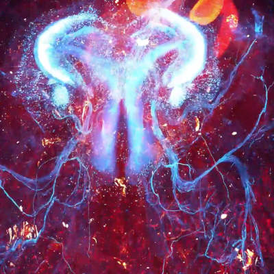

Scientists from the University of Science and Technology of China and other institutions in the Chinese Academy of Sciences have created a fast technology for high-resolution three-dimensional imaging of an entire small animal body. This imaging reaches subcellular resolution. The technology focuses on the peripheral nervous system, or PNS, which is the network of nerves outside the brain and spinal cord that connects the central nervous system to the rest of the body.
Traditional methods for studying the PNS used low-resolution anatomical studies at millimeter scale, which is about the thickness of a coin. Recent optical microscopy advances allowed micron-resolution imaging for whole brains, but these were too slow or not suitable for the larger, varied tissues of an entire body.
Adapting for Whole-Body Challenges
To overcome limitations for whole-body imaging, the researchers introduced an "in situ sectioning plus 3D blockface imaging" approach. In situ means done in place without moving the sample much. They built a system that combines a precision vibratome, a tool for slicing thin tissue layers, with the ARCHmap protocol, a method to make tissues transparent and embed them in hydrogel.
The system images a 600-micrometer-deep layer of the tissue surface in three dimensions, then removes a 400-micrometer-thick layer, repeating this cycle until the whole sample is processed. Automated algorithms stitch the images together using 200-micrometer overlaps for seamless alignment. This shallow depth reduces light scattering, where light bounces off uneven tissues, ensuring clear high-resolution results.
Using this pipeline, the researchers imaged an adult mouse body at uniform subcellular resolution in just 40 hours, producing about 70 terabytes of data per fluorescence channel, which detects glowing labels in samples. The method preserves fluorescent signals well and works with common neuroscience labeling techniques.
Through these images, the researchers examined detailed structures and paths of peripheral nerves. This advances PNS connectivity mapping and neural regulation understanding. Future improvements include multi-camera setups for faster multi-channel imaging and applications to larger biological samples. The breakthrough aids fields like developmental biology, comparative anatomy, and biomedical research by providing detailed data at population and single-cell levels.
This research is published in Cell.