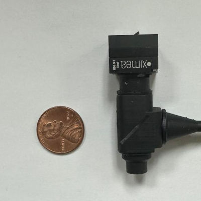

Scientists have created a small, light microscope that records the quick electrical signals from neurons in animals that are awake and moving freely.
his device weighs just over 16 grams and fits on the head of small animals like mice or rats. It looks through a tiny window in the skull to watch fluorescent dyes, which are special markers that glow brighter when neurons become active. Unlike older microscopes that track slower changes in calcium inside cells, this one catches voltage signals at up to 500 pictures per second. This speed lets it spot the exact moment a neuron fires, along with the small build-up signals before that happens.
The microscope uses a custom lens system with a high numerical aperture, a measure of how much light it can gather, to detect faint glow changes clearly. It pairs with a new dye called Voltron2 that responds strongly to voltage shifts without fading quickly. Tests on mice showed it matches the quality of larger lab microscopes, with clear spikes in signals that stand out from background noise.
Capturing detailed brain activity
By recording these fast voltage patterns in different brain areas, the tool helps reveal how neural circuits control actions like moving or thinking. For example, it could show how the hippocampus, a brain part for memory and navigation, times its signals. This might lead to better ways to treat brain disorders. The device has a small view area of 250 microns, about the width of a few hairs, but plans aim to make it lighter for more animal types and widen the view.
Overall, this advance offers a clearer picture of brain function in real-life settings, without heavy equipment limiting animal movement.
The scientists have described the methods and results of this study in a paper published in Biomedical Optics Express.