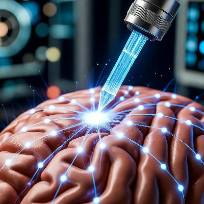

Existing tools called Neuropixels probes use high-density silicon arrays to gather electrophysiological data from the brain. These probes have helped collect data from animals like fish, reptiles, rodents, primates, and humans. They record from hundreds to thousands of neurons at once.
However, older Neuropixel probes cannot target tiny brain structures well and have trouble distinguishing electrical fields around separate cells.
The new Neuropixels Ultra probe solves some of these issues. It acts like an implantable camera that senses voltage to create flat images of a brain cell's electrical field. Voltage is the pressure that pushes electric current. The probe has much smaller and denser recording sites than past designs.
This work, published in Neuron, is part of the BRAIN Initiative, a program to create new tools for mapping brain circuits and understanding their roles. Brain circuits are connected groups of neurons.
Advantages of the new probe
In mouse tests, the probe recorded from the visual cortex, the brain area that processes eye input. It detected twice as many cells as older Neuropixels versions and separated three subtypes of cortical cells from others. This helps study brain circuits better.
The probe has higher noise per channel, but its many sites provide superior data quality. It excels at locating spikes, sudden electrical pulses from neurons, and keeping them separate from nearby cells. This precision boosts the number of usable neurons responsive to visual inputs.
Tests on other animals, including electric fish, bearded dragon lizards, and pigtailed macaques, showed the probe consistently finds small electrical patterns missed by lower-density tools. These patterns vary by genetically identified cell types, aiding in spotting cells and their activity.
The new probe trades some vertical coverage for high resolution. Older probes span more height in the brain. Still, the advance supports neuroscience by offering better brain activity details.