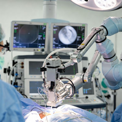

Surgeons at Boston University have shown that robotic surgery can safely remove the caudate lobe, a small and deep part of the liver that is hard to reach. This area is surrounded by important blood vessels and bile ducts. Removing tumors here is challenging because of the risk to these structures. The study, published in Annals of Surgical Oncology, focused on a 79-year-old patient with rectal cancer that had spread to the liver's caudate lobe. Using a surgical robot, the surgeons removed the tumor completely while protecting nearby vessels and ducts.
The procedure used two main tools for guidance. First, they applied a hanging method with the Arantius ligament, a thin band of tissue in the liver, to pull and create space for safe cutting near key vessels. Second, they used indocyanine green negative staining. Indocyanine green is a safe dye that glows under near-infrared light. They injected a small amount of the dye after blocking a blood flow branch to the caudate lobe. This made the rest of the liver glow while the caudate stayed dark, clearly showing the boundaries for removal.
Intraoperative ultrasound, which uses sound waves during surgery to create images of inside the body, helped map the tumor and vessels in real time.
Benefits of robotic approach
After the liver part, the surgeons also removed the main rectal cancer using the robot, making the whole operation minimally invasive, meaning it uses small incisions instead of large ones. The patient recovered without problems and finished further treatment. This method reduces pain, blood loss, and healing time compared to traditional open surgery. It aims to make complex liver operations safer and more accessible, especially for tumors in tough spots. The findings, published in a medical journal, suggest robotic precision combined with imaging tools could help more patients get curative surgery with fewer risks. Overall, this advances treatment for liver metastases, which are cancers that spread from other body parts.